
An error doesn't become a mistake until you refuse to correct it. ~ Orlando A. Battista
A while ago, a PET scan report landed on my desk. It was
a six-page document, detailing the methodology, the operative procedure with
organ-by-organ detailed results. All in all, it was a very impressive
presentation. The conclusion however after all the words suggested that the
results were negative; in other words the patient so tested failed to show any
abnormal activity in any of the organs.
“Good” I said and picked up the receiver and called to
inform the patient, which made her happy too. But there was this lingering dark
cloud turgid with contrary thought that lurked over my head. I pulled out the
previous report and compared them. Hmm. There was a discrepancy in the followup diagnostic testing somewhere and I, for my patient's sake, needed to find that out.
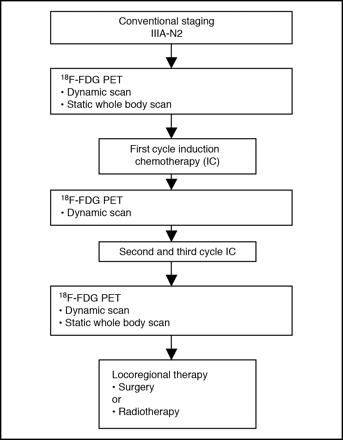
Follow up Algorithm of patient care
Sleuthing is a strange vocation. It keeps opening new doors
and closing others. The rabbit hole of progress takes you through some very
deep and dark alleys.
What is PET scan?
PET stands for Positron Emission Tomography: The positron is
a positively charged ion, the emission indicates a radioactive decay that is
being witnessed and captured by the sensors and the tomography is the detailed
radiological view of multiple planes of the human body. The single plane
technology was created by Robertson and colleagues in the Brookhaven Lab in
1961. Following that Wolf and associates discovered the 18F-FDG (Fluorodeoxy-D Glucose) for use as a radiopharmaceutical
as a scanned material in 1968. Abbas Alvi in University of Pennsylvania was the
first to use the 18F-FDG in normal human
volunteers for the first time in 1972.

Emissions: Signal to Noise Ratio
Let us focus on the emission aspect of this wonderful new
technology for all its worth. A
radiopharmaceutical agent is a radio-nucleotide that by virtue of their
instability undergoing decay leading to gamma emissions, which are captured by
the scanner. These gamma emissions from the sites that accumulate based on
oxygenation needs, occur at such a rapid pace that detectors with less than 10
nanosecond rate get very poor results due to the inability to resolve the “signal
to noise ratio.” Thus the results are sub-par and a lot of interpolation and
guesswork is involved. While newer machines with capabilities in the 100
picoseconds will have crisp data, it will be almost like comparing the new
digital OLED TV screens to the cathode ray tube of the yesteryear when these
new machines come online.
18F-Fluoro
Deoxy-Glucose and the Periodic Table
In the Periodic Table, Oxygen is the 8th element
and is followed by Fluorine, which is number nine. So the concept of creating
an 18-Fluorine from the 18-Oxygen arose initially by using
electrochemical fluoridation. Nowadays this is done by means of a Cyclotron,
bombarding protons at the 18-oxygen ions
in a 18-Oxygen “enriched water” and with a
“knockout reaction” displacing it with 18-Fluorine
ions. The 18-Fluorine ions have a
half-life of 109.8 minutes or under two hours.

FDG and the cell
Since the 18F-FDG is an
analog of the 18-Glucose the metabolically
active cells take it up for energy production. The phosphorylation of the 18F-FDG ~> 18F-FDG-6-Phosphate
prevents release of the glucose out of the cell. Since 18F-FDG is missing the 2’OH (Hydroxyl group) it is also unable to
be utilized in the glycolysis (glucose breakdown). This combined inability of
phosphorylation and non-utilization leads to an accumulation of the 18F-FDG-6P within the cell, thus represents the
glucose requirement of a functionally active cell as is seen in the normal
brain and the kidney and in the “high-octane” cancer cells.

PET scan result
18F-FDG
Metabolism and Excretion
The normal radioactive decay of 18F-FDG
yields 18-Oxygen-deoxyglucose, which picks
up an H+ from the hydronium ion in the liquid medium of the cell, thus creating
an OH (hydroxyl) group and a non-radioactive trans mutated 18O-Glucose-6 Phosphate that remains in the
cell, which is then metabolized through the usual pathway.

18F-FDG half life
Even though the half-life of 18F-FDG is 109.8 minutes, the disposal however is via two methods. 1) 75% is by
metabolism as described above into a harmless non-radioactive metabolite and 2)
25% is via direct kidney excretion in its radioactive mode (rapid elimination
prevents the half-life decay) in the form of radioactive urine excreted by the
patient. However, within 24 hours (13 half-lives), the radioactivity in the
patient and in any initially voided urine which may have contaminated bedding
or objects after the PET exam, will have decayed to 2^−13 = 1/8192 of the
initial radioactivity of the dose. So it is imperative to be careful of the
radioactive waste for at least 48 hours.
Okay now that we have figured this out, let me take you
deeper into the puzzle posed above in that PET scan report:
18F-FDG Half-Life and
Transportation
Given the half-life of 18F-FDg
is 109.8 minutes or under 2 hours, it means a facility without an in-house
cyclotron to create the 18F-FDG would have
to import such from another facility. That transportation time then has to be
incorporated into the value of the half-life. In other words if the transport
of the 18F-FDg took 2 hours then more than
half of the 18F-FDg would have decayed and
rendered useless, correct? So the game is to estimate into the transport system
the time lag of the transportation and send a larger dose that when it reaches
the facility will have enough volume and still be optimally radioactive and
capable of appropriate use. This is done via specially designed and regulated
transportation services. A further hitch would be the time the
radiopharmaceutical agent arrives at the hospital or facility and the
radiopharmaceutical-pharmacist accounts for it and then it gets transported to
the patient room and is dripped through the IV infusion into the patient.
By now you have guessed my confusion of that PET scan result
that I had obtained. Indeed the uptake was normal because not enough of the
functional 18F-FDG remained to give a
valid test. Repeating it with more stringent criteria revealed the error.
Pitfalls and other considerations in PET scanning
Another problem that sometime might happen is if there are
two metastatic cancer sites in an organ. One may have cells in an active state
of division, that site will necessarily uptake the majority of the 18F-FDG leaving little for the other less
functionally active site and therefore the PET results may show a high
intensity uptake or SUV (standardized uptake value) in the small tumor and a
weak signal uptake in the larger one, even though both are malignantly active.
The size of the tumors are better judged by a CT or an MRI scans and nowadays
the images of the PET can be merged with the CT and the MRI for better volume
delineation of the state of the human disease.

PET/CT scanner
As might be obvious, a high blood sugar value will minimize
the uptake of 18F-FDG since 18-F and 18-O
are “kissing cousins” on the periodic table, the high limit of blood sugar has
to be maintained below 180 mg/DL in order for the test to be performed
accurately. Additionally an active source of infection or chronic inflammation
will garner a major share of the 18F-FDG,
due to the glucose needs of the infected/inflamed sites. The differentiation
between the myriad issues that surround PET scanning is the purview of the
physician.
PETs are PETs but they still need to be patted on the head and deloused occasionally.


No comments:
Post a Comment